Interesting insect architecture
Editor’s note: This article is from the archives of the MSU Crop Advisory Team Alerts. Check the label of any pesticide referenced to ensure your use is included.
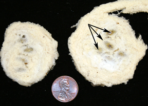
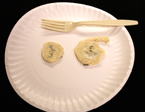
Last week a woman brought in some odd looking things that her carpenters found when they were removing some aluminum fascia from her roof. These things appeared to be concentric wrappings of insulation fibers about two inches in diameter. Located in the middle of the wrappings were some kind of insect pupal cases. My first guess was the pupae were those of a sphecid wasp. Sphecid wasps are well known builders of interesting nests. The organ-pipe mud dauber is a fine example.
In past years, several people have brought in sphecid pupae that they found crammed into the gaps between their window frames and siding. These pupae were enclosed individually in wrappings made of dried grass. These new nests were different from anything I had seen before.
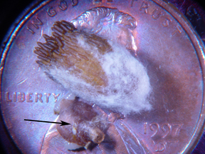
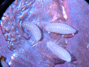
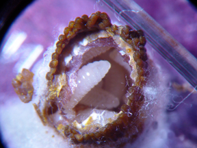
I removed a pupal case from the wrapping in hopes of finding a larva or pupa inside that would confirm my tentative identification. When I put the pupa under the scope, I saw that the mother wasp had carefully glued fecal pellets to the exterior of the pupal case, probably in an attempt to protect the pupae from parasitoids (others wasps that lay their eggs in or on another insect). I assumed the fecal pellets where from the developing sphecid wasp larva. When I dissected the pupa, I found that it had been parasitize by another wasp species. Instead of finding a single large pupa inside the case, I found a dozen of more parasitoid larvae. Apparently, the fecal pellets did not dissuade the parasitoid mother from laying her eggs inside the pupal case.
I am hoping to rear out both the sphecid and parasitoids to the adult stage. I’ll be sure to share the photos as they become available.


 Print
Print Email
Email